Services

EnquBio's Comprehensive Drug Discovery Solutions & Services

At EnquBio, our expert team delivers high quality, efficient, and dependable R&D solutions that enable pharmaceutical and biotech partners to advance and expand their drug development pipelines.

Therapeutic Areas · Inflammation & Autoimmunity · Respiratory

Human Airway ALI Model

A differentiated NHBE air-liquid interface epithelium with a validated Type 2 (IL-4 / IL-13) response, for asthma, COPD and respiratory drug discovery.

Standard 2D culture does not reproduce the human airway. EnquBio grows normal human bronchial epithelial (NHBE) cells at the air-liquid interface until they form a mature, pseudostratified mucociliary epithelium, so you can test compounds on the cell types and architecture your drug will actually meet.

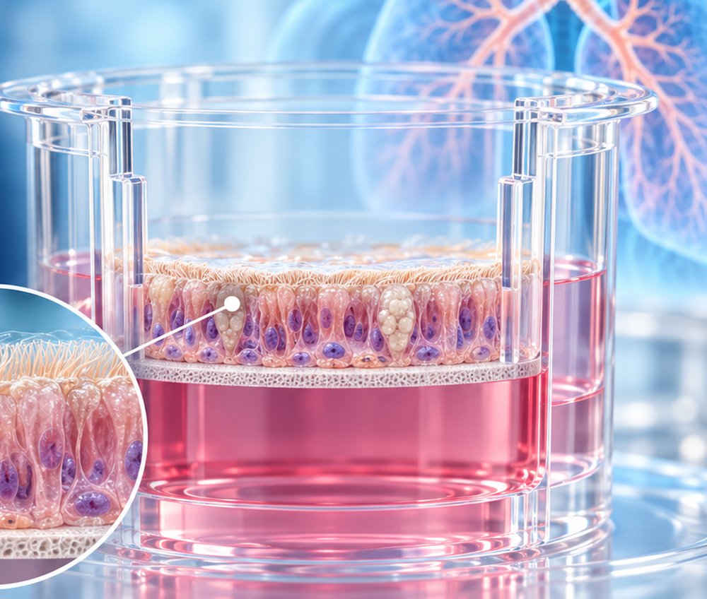
NHBE cells differentiated on a culture insert at the air-liquid interface
Cell sourcePrimary human NHBE
Mature epitheliumDay 28
Stable in cultureUp to 2 months
Type 2 responseIL-4 / IL-13 validated

How the air-liquid interface rebuilds the airway

Cells sit on a porous membrane. Air reaches the apical surface while medium feeds the basal side, the same orientation as the bronchial lining. That polarity is the signal that drives differentiation.

Air-liquid interface culture, cross-section NHBE cells grow on a porous membrane. The apical surface faces air; medium feeds the cells from the basal side. The cells form a pseudostratified epithelium of basal, ciliated and goblet cells. AIR MEDIUM Mucus layer secreted onto the apical surface Ciliated cells acetyl-α-tubulin+ Goblet cells MUC5AC+, mucus-producing Basal cells progenitor layer Porous membrane insert support Basal medium feeds cells from below

Validated mucociliary differentiation

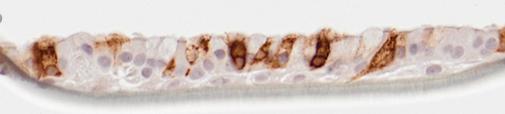
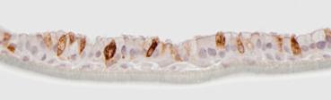
EnquBio immunohistochemistry at Day 28. Select a marker to view the stained sections.

MUC5AC immunohistochemistry, NHBE ALI section, Day 28 MUC5AC immunohistochemistry, second field

Representative sections, Day 28 post-airlift. Brown: positive staining.

Mucus-producing cells

MUC5AC+ goblet cells

MUC5AC marks the mucin-secreting goblet cells. Their presence confirms the secretory lineage has differentiated and that the culture can model mucus biology.

  • Pseudostratified architectureBasal, ciliated and goblet cells in one layered epithelium
  • Long-term stabilityDifferentiated phenotype maintained for up to 2 months
  • Primary human cellsDonor-derived NHBE, not an immortalized line

Type 2 airway response: IL-4 / IL-13 stimulation data

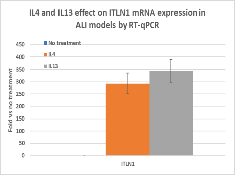
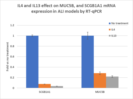
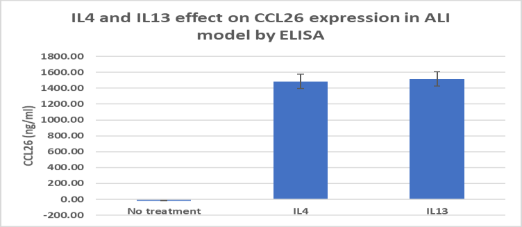
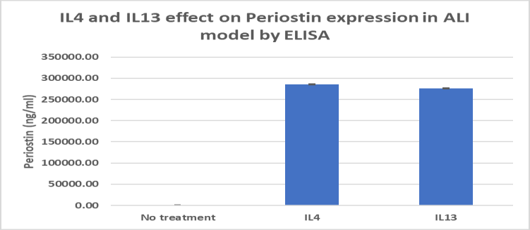
Five readouts from EnquBio's stimulated NHBE ALI cultures. Charts are redrawn from the brochure figures; the original figure is one click away.

Assay

Readout

Gene expression · RT-qPCR

MUC5AC & SPDEF

Goblet-cell and mucus genes

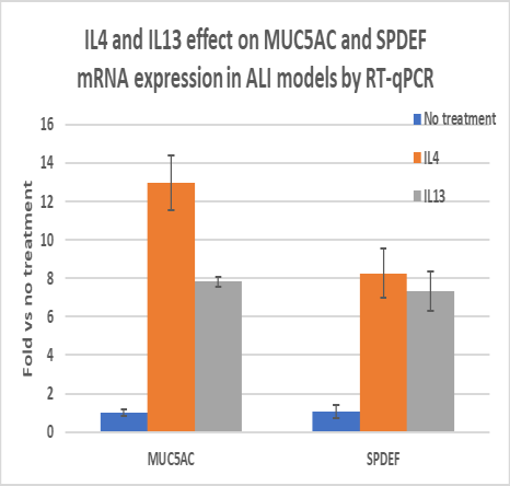
MUC5AC and SPDEF mRNA, RT-qPCR 0 4 8 12 16 Fold vs no treatment MUC5AC SPDEF No treatment IL-4 IL-13

IL-4 and IL-13 each increased MUC5AC and SPDEF relative to untreated cultures, consistent with Type 2-driven goblet-cell differentiation.

Source: EnquBio NHBE ALI Model brochure. n and statistics as in source. Compound-mediated inhibition of these responses is study-specific and not shown here.

Why choose ALI over conventional 2D culture

The question is whether your readout depends on airway architecture. If it involves mucus, cilia, barrier polarity or apical exposure, a submerged monolayer cannot answer it.

Submerged 2D / immortalized linesEnquBio NHBE ALI
CellsOften transformed lines (e.g. BEAS-2B)Primary normal human bronchial epithelial cells
ArchitectureSingle flat layerPseudostratified epithelium
Goblet cells / mucusAbsentMUC5AC+ goblet cells
Ciliated cellsAbsentAcetyl-α-tubulin+ ciliated cells
Type 2 responseLimited; no goblet-cell metaplasiaIL-4 / IL-13 induces MUC5AC, ITLN1, CCL26, periostin
Apical exposureCompound diluted in mediumApical surface open to air for topical or inhaled-type dosing
Study windowTypically 24–48 hStable for up to 2 months

Disease applications

One application is backed by the data above. The others are built as custom studies after a feasibility discussion.

Demonstrated response

Asthma & Type 2 airway inflammation

IL-4 / IL-13 stimulation reproduces the goblet-cell, mucin and chemokine signature of Type 2 asthma (MUC5AC, SPDEF, ITLN1, CCL26, periostin). Use it to test whether your compound blocks that response in primary human airway epithelium.

Custom study scope

COPD & epithelial dysfunction

Mucociliary dysfunction and epithelial inflammation in a differentiated human airway.

Custom study scope

Cystic fibrosis

Mucus biology on a human mucociliary epithelium.

Custom study scope

Respiratory infection

Apical application of pathogens or mimetics, the natural route of airway exposure.

Custom study scope

Inhalation & drug delivery

Apical dosing to assess local epithelial effects of inhaled and topical formulations.

Custom study scope

Inflammatory mechanisms

Cytokine, chemokine and remodeling-marker readouts linked to your target.

Readouts

The three used in the validation data, plus the EnquBio platforms an ALI study can draw on.

  • RT-qPCRGene expression: mucins, Type 2 response and differentiation markers
  • ELISASecreted cytokines, chemokines and remodeling proteins from apical wash or basal medium
  • ImmunohistochemistryGoblet and ciliated cell markers, epithelial architecture
  • MSD multiplexMultiplexed cytokine panels on the same supernatants
  • Western blot / JessPathway activation in epithelial lysates
  • Custom pharmacologyCompound, dose, timing and collection schedule designed per study

Frequently asked questions

What is an air-liquid interface (ALI) airway model?

Primary human bronchial epithelial cells are grown on a porous membrane with air above and culture medium below. Air exposure drives them to differentiate into a pseudostratified epithelium with mucus-producing goblet cells and ciliated cells, closely resembling the human airway lining.

Why primary NHBE cells instead of an immortalized line such as BEAS-2B?

Primary NHBE cells retain the ability to differentiate into goblet and ciliated cells at the air-liquid interface. Immortalized lines grown submerged do not form a mucociliary epithelium, so they cannot report on mucus production, ciliated-cell biology or epithelial architecture.

How long does differentiation take, and how long are cultures stable?

Cultures form a mature pseudostratified epithelium by Day 28 and remain stable in vitro for up to 2 months, confirmed by immunohistochemistry.

Has the model been validated for Type 2 (asthma) inflammation?

Yes. IL-4 and IL-13 stimulation increased MUC5AC, SPDEF and ITLN1 expression, decreased SCGB1A1 and MUC5B, and increased CCL26 and periostin secretion, a characteristic Type 2 airway response.

Is ALI work performed under GLP?

No. ALI studies run as non-GLP discovery and translational research, which keeps design flexible and turnaround fast for target validation, compound ranking and mechanism-of-action work.

Related services

Planning a respiratory study?

Tell us your target, indication and the readout that matters. Our scientists will propose an ALI study design and timeline.

Contact Our Scientists →